How does NMR spectroscopy work?
NMR spectroscopy relies on some tricky concepts, but the process itself is relatively simple. You follow these steps:
- Dissolve your sample in a suitable solvent, such as .
- Add a small amount of a reference molecule, such as TMS.
- Place the sample in an external magnetic field.
- Fire radio waves at the sample.
NMR spectroscopy, short for nuclear magnetic resonance spectroscopy, is an analytical technique we use primarily to find out the structure of molecules. It is based on the behaviour of certain nuclei in an external magnetic field.
Nuclei in the sample absorb and emit radio waves according to the other atoms or groups bonded to them. These waves are detected by a detector. The detector produces a spectrum showing the energy absorbed against a property called chemical shift.
What is chemical shift?
You’ll learn more about the science behind NMR spectroscopy, including chemical shift, in Understanding NMR. However, we’ll take a quick look at it now to help you understand how NMR spectroscopy works.
As we mentioned, NMR spectra show the chemical shift of nuclei. This is a property related to something called magnetic resonance frequency. Chemical shift is measured in parts per million, or ppm.
To summarise briefly, certain nuclei act a little weirdly when placed in an external magnetic field. They take one of two states: parallel, known as spin-aligned; or antiparallel, known as spin-opposed. If we supply them with enough energy they can flip from their parallel to their antiparallel state. This energy is called their magnetic resonance frequency.
Magnetic resonance frequency is the energy needed for a nucleus to flip from its parallel to its antiparallel state in an external magnetic field.
Magnetic resonance frequency varies depending on the environment of an atom.
An atom’s environment is all the different chemical groups attached to it.
Identical nuclei from the same element can have different magnetic resonance frequencies and different chemical shift values if they are bonded to different groups, because they are found in different environments. This is the fundamental concept behind NMR spectroscopy.
Only nuclei with odd mass numbers can be used in NMR spectroscopy. This is because they have a property called spin. You'll learn more about spin in Understanding NMR.

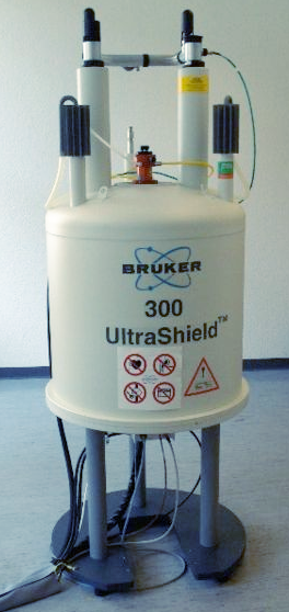
A high power NMR spectrometer,
Andel Früh & Andreas Maccagnan, CC BY-SA 3.0, via Wikimedia Commons [1]
Interpreting NMR spectra
As we mentioned above, NMR spectroscopy produces graphs called spectra, plotting energy absorbed by the sample against chemical shift. The graphs show a number of different peaks. Nuclei from identical atoms produce peaks at different chemical shift values depending on the other atoms or groups of atoms bonded to them. Notice the peak shown at 0 ppm. This is given by TMS, a reference molecule.
Tetramethylsilane, also known as TMS, is a molecule commonly used as a reference point in NMR spectroscopy.
 An example of an NMR spectrum, showing distinct peaks. Note the peak given at 0 ppm by TMS, the reference molecule. Anna Brewer, StudySmarter Originals
There are two important things to know:
- Environments with certain functional groups produce chemical shift peaks that fall within a particular range.
- Unique environments give unique chemical shift peaks.
How does this help us? Well, if you have two clear peaks on your spectrum, your sample must contain nuclei in two different environments. You can then compare the chemical shift value of the peaks to values in a data book, which will tell you what sort of environment the nuclei are in, and the different functional groups that are attached to them. This helps you work out the structure of the molecule in your sample.
Let’s say you have the following spectrum for an unknown molecule.
 A carbon-13 NMR spectrum. Anna Brewer, StudySmarter Originals
You can see peaks at around 58 ppm, 18 ppm and 9 ppm. Let’s compare these values to a data table.
 A typical data table for carbon-13 NMR. Anna Brewer, StudySmarter Originals
The peak at 58 ppm matches the values for an group, which range from 50-90 ppm. We can therefore infer that this molecule contains that particular group. Similarly, we can see that the peak at 18 ppm falls into the range for an group, and the peak at9 ppm falls into the range for an group.
What molecule do you know that contains just these particular groups? Let’s put them together:
 Our mystery molecule is propan-1-ol. Anna Brewer, StudySmarter Originals
The molecule is propan-1-ol.
In summary, by comparing chemical shift values to ranges in a data book, we can infer the different groups within a molecule and work out its overall structure.
Different types of NMR spectroscopy
Not all nuclei can be used in NMR spectroscopy. Most aren’t influenced by an external magnetic field and can’t be detected. Two types of nuclei that do produce results in NMR spectroscopy are carbon-13 nuclei and hydrogen-1 nuclei.
Remember that carbon-13 shows that we have an isotope of carbon with a mass number of 13. Mass number is the combined number of protons and neutrons in an atom. Carbon has an atomic number of 6, meaning it has six protons, and so carbon-13 atoms must have 13 - 6 = 7 neutrons.
Both types of spectroscopy follow the general technique described above and detect the chemical shift of carbon-13 nuclei and hydrogen-1 nuclei respectively. However, the chemical shift peaks in hydrogen-1 spectra fall within a much smaller range.
Hydrogen-1 NMR spectroscopy is also known as proton spectroscopy. A hydrogen-1 nucleus doesn’t have any neutrons or electrons - it is just a proton.
 A hydrogen-1 atom. If you take away the electron you are left with only the nucleus, which contains just one proton. Anna Brewer, StudySmarter Originals
Hydrogen-1 NMR spectroscopy does have some advantages over carbon-13 spectroscopy:
- Most hydrogen atoms are the isotope hydrogen-1, whereas only about 10 percent of carbon atoms are the isotope carbon-13. This means that hydrogen-1 spectra give clearer, more distinct results.
- The size of peaks in hydrogen-1 spectra is proportional to the number of hydrogen-1 nuclei in that particular environment, which isn’t the case for carbon-13 NMR peaks. This is shown using an integration trace.
- Hydrogen-1 peaks show something called spin-spin coupling. This is where they split into smaller peaks depending on how many hydrogen atoms are in adjacent environments, and it gives us further information about the molecule's structure.
 This hydrogen-1 spectrum for ethanol shows spin-spin coupling. Some peaks have split into multiple smaller peaks. Anna Brewer, StudySmarter Originals
You’ll learn more about carbon-13 and hydrogen-1 NMR in Carbon -13 NMR and Hydrogen -1 NMR respectively.
Uses of NMR spectroscopy
NMR spectroscopy has many applications in modern science. As we’ve explored, its primary function is analysing molecule structure and shape. However, it is also used for the following purposes:
- Determining protein folding.
- Drug screening and design.
- Finding out how molecules interact in chemical reactions.
- Determining the proportion of solids and liquids in lipids.
Pros and cons of NMR spectroscopy
NMR Spectroscopy has both pros and cons. Let's consider them below.


